Figure 6.
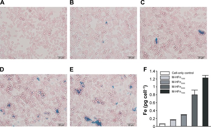
Iron uptake analysis of MDA-MB-231 tumor cells incubated with M-HFn nanoparticles.
Notes: Prussian blue staining of MDA-MB-231 tumor cells incubated for 24 hours with (A) no nanoparticles, (B) M-HFn1000, (C) M-HFn3000, (D) M-HFn5000, and (E) M-HFn7000. (F) Iron contents in single cell are 0.16 pg cell−1, 0.29 pg cell−1, 0.80 pg cell−1, and 1.23 pg cell−1 after incubation with M-HFn1000, M-HFn3000, M-HFn5000, and M-HFn7000, respectively, for 24 hours (statistical comparison of iron contents in single cell with cell-only yielded P=0.014, 0.002, 0.011, and 0.023 for M-HFn1000, M-HFn3000, M-HFn5000, and M-HFn7000, respectively).
Abbreviation: M-HFn, ferrimagnetic H-ferritin.

