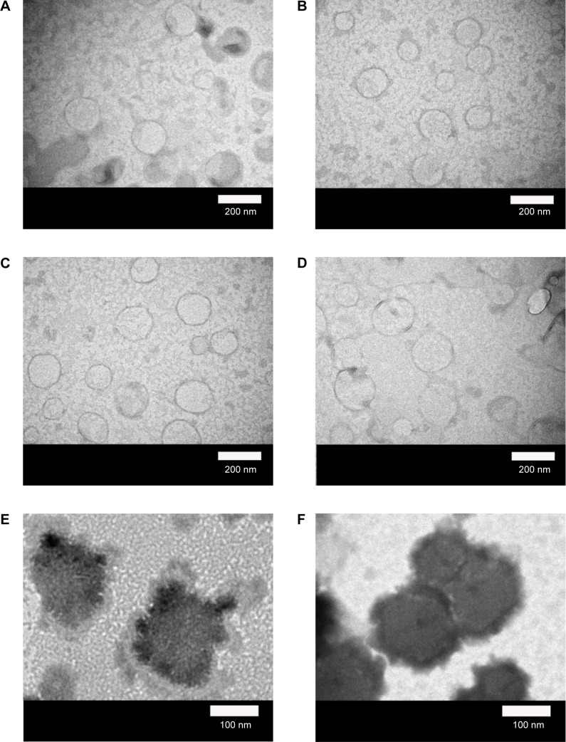
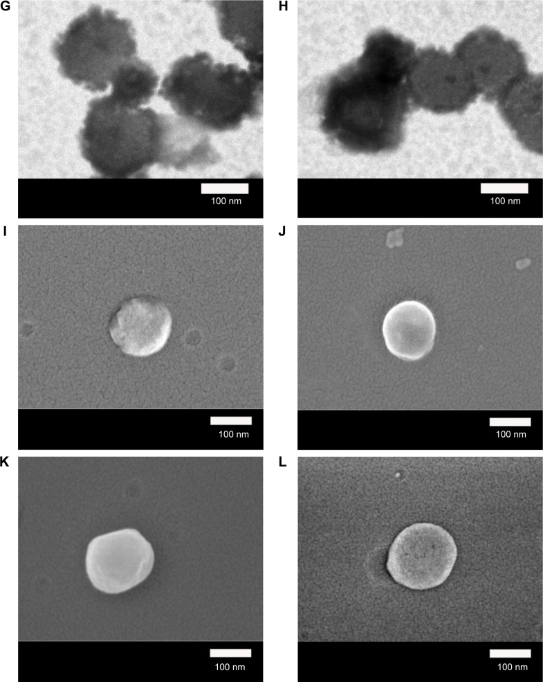
Figure 3.
Micrographs of WGA-CL-NGF-CUR-liposomes.
Notes: (A–H) Transmission electron microscopic images. (I–L) Scanning electron microscopic images. (A, I) rCL =0%; (B, J) rCL =5%; (C, K) rCL =10%; (D, L) rCL =20%; (E) CWGA =2.5 mg/mL and rCL =10%; (F) CWGA =2.5 mg/mL and rCL =20%; (G) CWGA =5 mg/mL and rCL =10%; (H) CWGA =5 mg/mL and rCL =20%.
Abbreviations: rCL, CL mole percentage in lipids (%); CWGA, WGA concentration in grafting medium (mg/mL); CL, cardiolipin; CUR, curcumin; NGF, nerve growth factor; WGA, wheat germ agglutinin.


