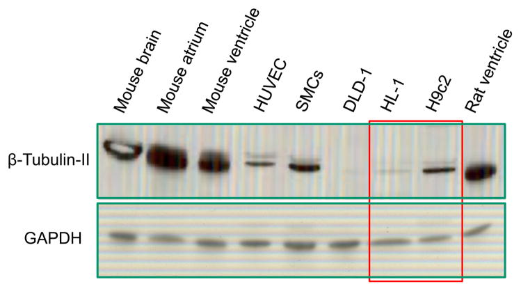
Fig. 3.
Western blot analysis of beta-tubulin II in various cells and tissues. Similarly to confirming the results of fluorescent confocal imaging (see Fig. 2), the presence of beta-tubulin II in H9c2 cells and the absence in HL-1 cells were found. Note the abundant amount of beta-tubulin II in rat and mice heart and in brain. All probes contained app. 20 μg of total protein. GAPDH (“Ambion”) was used a loading control. DLD, human colorectal carcinoma cells; HUVEC, human umbilical vein endothelial cells; SMS, smooth muscle cells.

