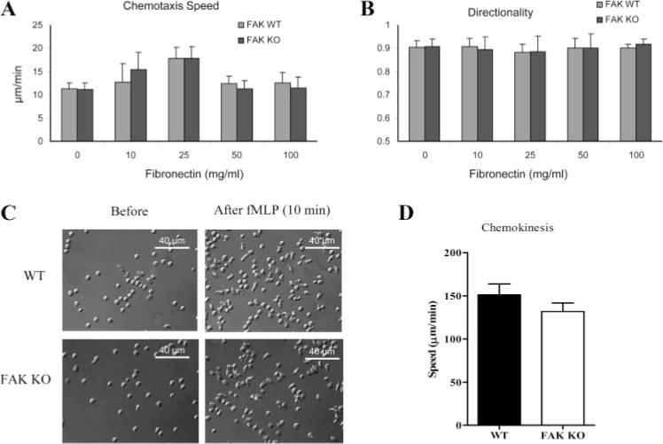
FIGURE 2.

FAK−/− neutrophils exhibit normal chemotaxis and chemokinesis upon fMLP stimulation. A and B, Wild-type (WT) and FAK−/− (KO) neutrophils were plated onto uncoated or fibronectin-coated coverslips. Chemotaxis was visualized using an EZ-TAXIScan device (supplemental Fig. 4). Chemotaxis speed and directionality were quantified using DIAS imaging software as described in supplemental Fig. 5. Results are presented as means ± SD of >20 neutrophils. C, Bone marrow-derived wild-type and FAK−/− neutrophils were plated onto fibronectin (10 μg/ml)-coated MatTek dish and uniformly stimulated with 100 nM fMLP. Shown are representative images of neutrophils before (left) and 10 min after fMLP stimulation (right). Two videos of the experiment described in this figure are included in supplemental Movie 6). Images were captured using a ×40 optical lens (recorded at 1 frame/10 s). D, Migration speeds of wild-type and FAK−/− neutrophils during chemokinesis were calculated as total distance of migration from origin divided by total time. Results are means ± SD of 10 neutrophils. ■, Wild type; □, FAK−/−.
