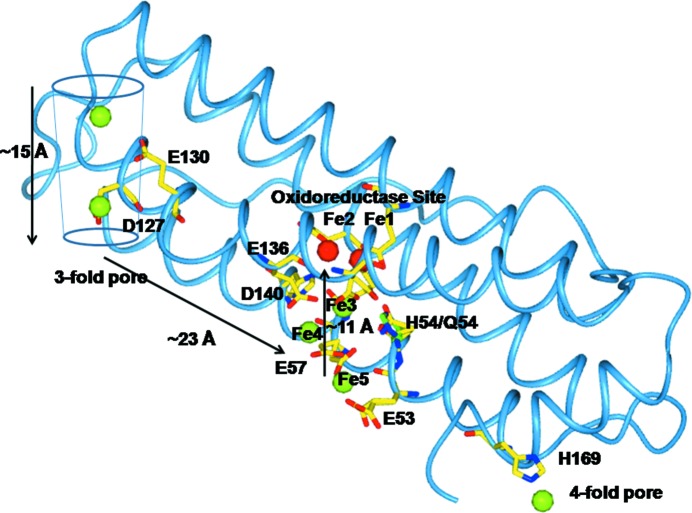
Figure 6.
The travel of Fe2+ ions inside the RcMf subunit. The probable pathway of Fe2+ ions of about 50 Å in length from the threefold axis channel to the oxidoreductase site as indicated by the crystallographic structures. The residues involved in Fe2+ binding (green spheres) are shown as sticks. The two Fe2+ ions bound to Fe1 and Fe2 sites are shown as red spheres. The Fe2+ ion trapped at the fourfold axis pore is shown as a green sphere.

