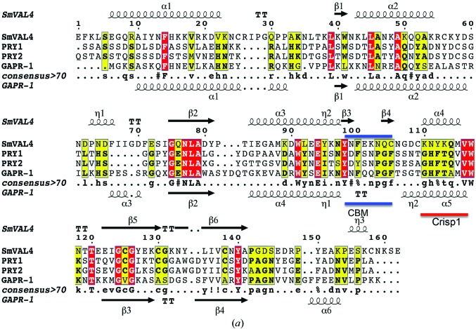
Figure 6.
The calveolin-binding motif. (a) The conserved calveolin-binding motif (CBM) is evident in the alignment of the sequences of SmVAL4, GAPR1, Pry1 and Pry2. The secondary-structural elements shown are for SmVAL4 and GAPR1 (PDB entry 4aiw). The location of the CBM is identified with a blue line, while the CRISP1 motif is shown as a red line.

