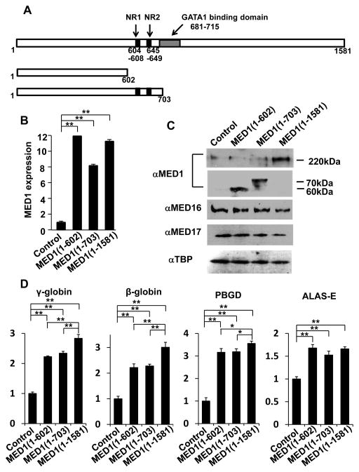
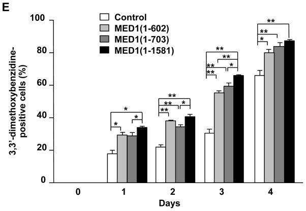
Figure 3. MED1(1–602) and MED1(1–703) induce GATA1-mediated transcription and erythroid differentiation in K562 cells.
(A) Diagram of MED1, showing the domains of 2 NR boxes (NR1 and NR2) and the GATA1 binding domain and the MED1 fragments used in these experiments.
(B, C) Results of quantitative RT-PCR for MED1 (B) and western blot analyses for Mediator subunits and TBP as a control (C) 3 days after transfection with expression vectors with various fragments of MED1 or an empty expression vector as a control in hemin-treated K562 cells. Values (mean ± SD) of a representative experiment performed in duplicate are shown (**p < .01) (B).
(D) Enhanced GATA1-targeted gene transcription after overexpression of various fragments of MED1 or an empty expression vector as a control in hemin-treated cells. Values (mean ± SD) of a representative experiment performed in quadruplicate are shown (*p < .05, **p < .01).
(E) Enhanced erythroid differentiation after overexpression of various MED1 fragments in hemin-treated cells. An empty expression vector was used as a control. Values (mean ± SD) of a representative experiment performed in triplicate are plotted (*p < .05, **p < .01).
The results were reproducible in three independent experiments.


