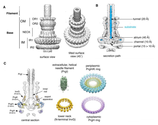
Fig. 1.
Needle complex structure from Salmonella typhimurium. A. Surface views of the 3D reconstruction of the cryo EM map of the S. typhimurium needle complex. The different substructures are noted. B. Surface view of a half-sectioned needle complex containing a trapped substrate within the central tunnel (119). Relevant structural details and dimensions are noted. C. Docking of the atomic structures of the different needle complex components onto the 3D cryo-EM map.

