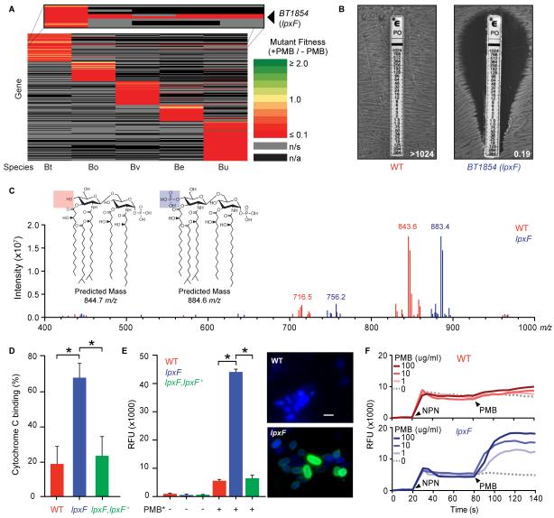
Fig. 2. Lipid A dephosphorylation mediates AMP resistance in prominent human gut bacteria.
(A) Heat map of the relative fitness of transposon mutant strains in the presence and absence of PMB. Columns indicate human gut bacterial species tested (abbreviations from Fig. 1B); rows indicate gene orthologs with insertions displaying a significantly altered fitness (q<0.05) in the presence of PMB in at least one species. n/s, not significantly altered; n/a, no ortholog. (B) BT1854 (LpxF) is necessary for PMB resistance in B. thetaiotaomicron. Representative MIC assessed using the E-test method is shown. (C) LpxF is required for dephosphorylation of lipid A. FT-ICR MS analysis of lipid A isolated from wildtype (red) and lpxF deletion mutant (blue) B. thetaiotaomicron revealed a shift of the predominant peak by ~40 m/z, consistent with the gain of a single phosphate group. Inset shows predicted structures and m/z values of the doubly deprotonated ([M-2H]2−) ions for monophosphorylated (red) and bis-phosphorylated (blue) penta-acylated lipid A. Minor peaks are consistent with tetra-acylated lipid A structures (fig. S2C-D). (D) Deletion of lpxF increases cationic cytochrome C binding to B. thetaiotaomicron, indicating altered surface charge. (E) Increased PMB-Oregon Green (PMB*) binding to lpxF deletion mutant cells measured by fluorescence quantification (left panel) and microscopy (right panels). Error bars represent standard deviation and asterisks indicate significance (p<0.01). Representative fluorescence microscopy images of bacterial cells incubated with PMB* (green) are shown as an overlay with 4′, 6′ diamino-2-phenylindole (DAPI) (blue). Scale bar indicates 2 μm. (F) LpxF protects the outer membrane from PMB perturbation. 1-N-phenylnaphthylamine (NPN) uptake profiles of select strains were measured followed by challenge with the indicated concentration of PMB. Arrowheads indicate the addition of NPN (20s) and PMB (80s). Readings were taken in 5s intervals. Each experiment was performed in triplicate with representative results shown. See figure S3 for complementation.

