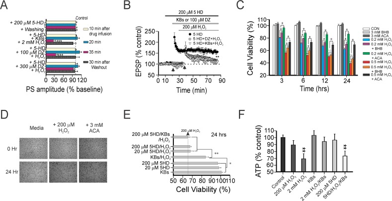
Fig 3. Pharmacological blockade of KATP channels negates the hippocampal synaptic and neuronal protection afforded by ketones.
(A) Changes in PS amplitude (as % of baseline) during or after drug application under different experimental conditions. No changes were seen in the PS amplitude during infusion with 200 μM 5HD alone or 5HD with 300 μM DZ and H2O2. There was a reversible, dose-dependent blockade by 5HD of the PS when either ketones or 100 μM DZ were applied concurrently with H2O2. Asterisks denote significant differences between experimental groups and the control at a specific time-point (*** p < 0.001). The dotted line indicates the mean PS amplitude of the control group. Each horizontal bar indicates mean PS amplitude ± SEM obtained in 12 slices from 5 rats. (B) Normal TBS-induced LTP was seen with 200 μM 5HD. However, LTP was impaired when either ketones or 100 μM DZ were administered together with 5-HD; the EPSP amplitude was measured 110 ± 9% and 115 ± 11% at 50 min post-TBS, respectively. Asterisks denote significant differences between 200 μM 5-HD alone and other treatment groups (**, p<0.01). Each LTP dataset was collected in 10 hippocampal slices. (C) Time-course of the MTT assay for cell viability in murine hippocampal HT22 cells treated with H2O2—with or without application of BHB or ACA (each 3 mM)—for 3, 6, 12, and 24 hours. Each vertical bar indicates the mean ± SEM (n = 30). (D) Representative photomicrographs of HT22 cells under control conditions, with H2O2, or when H2O2 was co-treated with ACA at two time-points (0 and 24 hrs). (E) Oxidative-induced cell death in HT22 cells measured under different treatment conditions. While 20 μM and 200 μM 5HD alone had no influence on cell viability, ketone (each 1 mM)-mediated neuronal protection against oxidative injury was completely negated by 200 μM 5HD. (F) Bar graphs illustrating changes in ATP levels in hippocampal CA1 samples treated with ketones with or without 5-HD on H2O2-induced oxidative stress after 2 hrs. ATP levels represented as % of control are mean ± SEM of 12 slices analyzed after 2 hr with indicated significant decreases (**p < 0.01) compared with control group, which was infused with physiological saline under similar experimental conditions.

