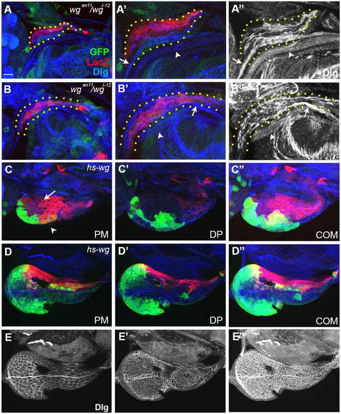
Fig 3. Wg is important for the growth of ead and the establishment of the ventral domain.
A and B were magnified 2 times in A’ and B’. Cell boundaries visualized with anti-Dlg antibody are shown in A”, B” and E. (A) wg I-12 / wg en-11 ead with no ventral domain. Bolwig’s nerve and the unknown nerve are marked with arrow and arrowhead, respectively. (B) An older ead from a wg I-12 / wg en-11 larva after prolonged culture. (C) hs-wg ead. (C”) is a combined image of (C’, PE) and (C, DP). (D, E) Some midline (arrow) and ventral wg-LacZ+ (arrowhead) cells are missing in hs-wg ead. (E-E”) The images of Dlg in (D-D”) show the cell boundary and Bolwig’s nerve. Scale bar, 10 μm.

