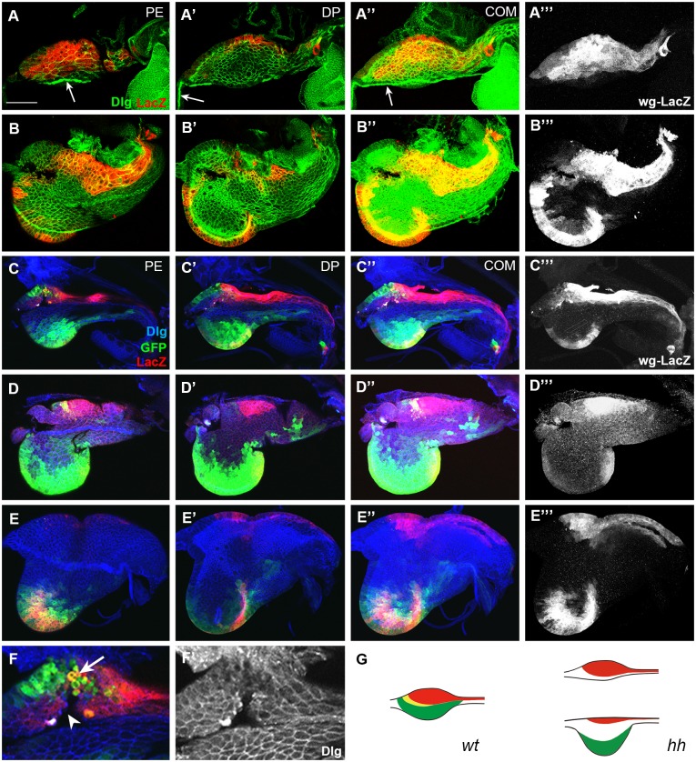
Fig 4. Domain-specific effects of hh mutation in eads.
(A) An early L2 hh ts2 disc without ventral domain. Bolwig’s nerve is marked with an arrow. (B) An early L3 ead with wg-LacZ+ cells at the entire ventral margin. (C) A late-L2 hh ts2 ead with small dorsal domain without midline cells. (D) An early L3 ead without dorsal posterior domain in contrast to large ventral domain. (E) A late L3 ead without any dorsal dpp-gal4+ cells and only a small number of dorsal wg-LacZ+ cells. (F) Magnified image of C showing the damaged dorsal tissue (arrowhead) with round-shaped cells (arrow). (G) A diagram of hh ts2 eads, one without the ventral domain, the other with large ventral domain. Scale bar: A, 20 μm; B, C, D, 30 μm; E, 100 μm; F, 10 μm.

