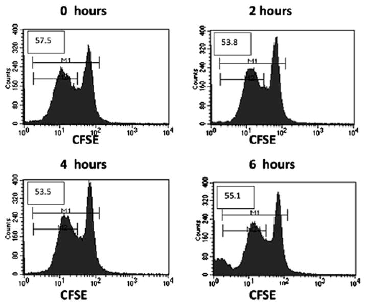
Fig. 2.
Addition of CPD to RBCs does not suppress T-cell proliferation within the first 6 h after exposure. We added fresh RBCs to anti-CD3/CD28 stimulation cultures of CFSE-labeled T cells after the indicated times of incubation with CPD. T cells were harvested after 3 d and proliferation was measured by CFSE dye dilution using flow cytometry. Initial CFSE-stained T-cell populations are demonstrated by the large peaks at a fluorescence intensity of 102; the leftward diminution of fluorescence intensity demonstrates continued proliferation. Numbers in each histogram represent the percentage of T-cell division (M2/M1 x 100).

