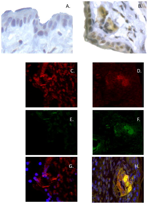
FIGURE 6.
Tissue localization of CXCL1 to endothelial cells during elicitation of CHS. BALB/c mice were sensitized with 0.25% DNFB on days 0 and +1. On day +5 after sensitization, mice were challenged on a shaved square area of trunk skin with 0.2% DNFB. Challenged areas of skin were removed at 6 h post-challenge from DNFB-sensitized or non-sensitized mice and (AB) Paraffin-embedded sections were prepared and stained with CXCL1-specific antiserum. Slides were examined by light microscopy and representative images from (A) naïve and (B) sensitized skin are shown. Magnification, 40×. C–H Frozen sections were prepared and stained with both CXCL1-specific antiserum and anti-CD31 antibody followed by fluorochrome-labeled secondary antibodies (red to react with the CD31 and green to react with the CXCL1-specific antiserum) and were examined by confocal microscopy. Representative images of CD31 (C and D), CXCL1 (E and F), or both CD31 and CXCL1 (G and H) staining in challenged skin from naïve (C, E, and G) and sensitized (D, F, and H) mice are shown. Magnification, 40×.

