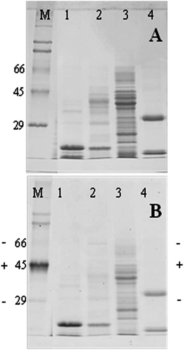
Fig 2. Detection of phosphoryl groups in Blad-oligomer.
Blad-oligomer, α, β, and γ-conglutins were purified from L. albus, subjected to SDS-PAGE, and stained for total protein (A) or analysed for the presence of phosphoryl group using the Pro-Q diamond phosphoprotein gel stain (B). Lanes 1, 2, 3, 4: Blad-oligomer, α, β, and γ-conglutins, respectively. Lanes M: molecular mass standards (kDa). The position of the positive (+) and negative (-) phosphorylated markers is shown in B.

