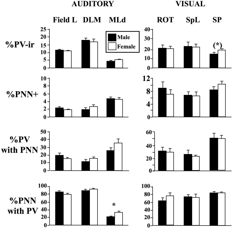
Fig 4. Sex differences in the percentage of DAPI cells that were immunoreactive for parvalbumin (%PV-ir cells) or were surrounded by perineuronal nets (%PNN cells), in the percentage of PV-ir cells surrounded by PNN (%PV with PNN) and in the percentage of PNN surrounding PV-ir cells (%PNN with PV) in 3 auditory areas (Field L; the dorsal lateral mesencephalic nucleus, MLd; the medial nucleus of the dorsolateral thalamus, DLM), and 3 visual areas (the nucleus rotondus, ROT; the nucleus spiriformis lateralis, SpL; the nucleus subpretectalis, SP).
The figure shows the mean ± SEM of data in males (black bars) and females (open bars). Numbers of data in each case are listed in Table 1. * = p<0.05 and (*) = 0.10<p<0.05.

