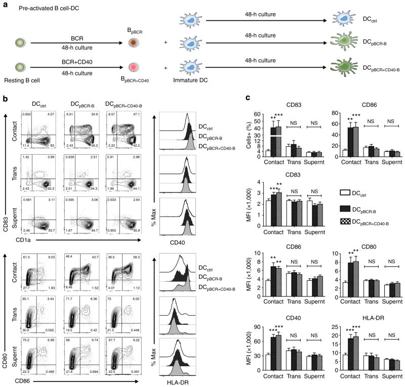
Figure 2. Cellular contact is required and sufficient for the induction of maturation of DCs by activated B cells.
(a) Pre-activated B cell–DC experimental design. Immature DCs were cultured in medium containing granulocyte macrophage colony-stimulating factor and IL-4 for 48 h either alone (DCctrl) or co-cultured at 1:1 ratio with CD19 + B cells that were pre-activated by BCR (DCpBCR-B) or BCR + CD40 (DCpBCR + CD40-B) stimulation. (b,c) DC–B cell co-culture was either done in 96 U-bottomed wells to allow direct contact between DCs and B cells (Contact) or in transwell plate to separate the B cells from DCs (Trans). Immature DCs were also cultured in the supernatants from activated B cells (Supernt). Phenotypic analysis (% positive cells and mean fluorescence intensity, MFI) of DCs that were gated negative for CD20. Representative plot and mean±s.e.m. of data from 7 to 11 donors. (Contact, n =11; Transwell, n =7; Supernatant, n =8). **P<0.01; ***P<0.001; NS, not significant by one-way analysis of variance test.

