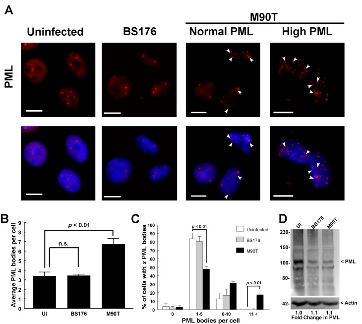
Fig 1. Shigella infection increases PML-NB number.
HeLa cells were not infected (UI), infected with a non-invasive strain (BS176), or infected with an invasive wild-type strain (M90T) as indicated, both strains contained a plasmid expressing the afaE adhesin (A). Cells were incubated for 3 hours, fixed and immunostained for PML (red). M90T-infected cells with normal and high levels of PML nuclear bodies are shown as indicated. DNA was visualized with DAPI (blue) and white arrow heads indicate PML NB “doublets”. Scale bars = 10μm. The average number of PML bodies per cell for each treatment condition was quantified (B) and the p-value of Student’s t-test was calculated (n.s. = not significant). The percentage of cells containing either 0, 1–5, 6–10 or 11+ PML bodies was also determined (C) and the p-value of Student’s t-test was calculated. Values in B and C represent the mean +/- SE, n = 3. Total PML protein levels for each treatment condition was determined by Western blot analysis (D).

