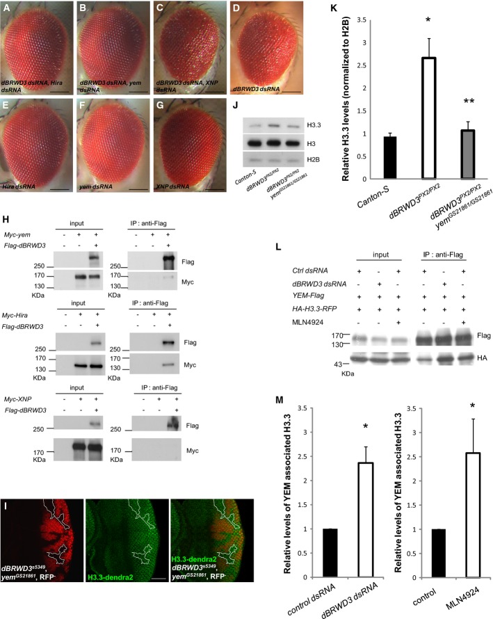
A-G Suppression of dBRWD3-RNAi induced rough eye phenotype by simultaneous Hira or yem depletion. Images of adult eyes: OK107-GAL4-driven UAS-dBRWD3-dsRNA, UAS-Hira-dsRNA (A), UAS-dBRWD3-dsRNA, UAS-yem-dsRNA (B), UAS-dBRWD3-dsRNA, UAS-XNP-dsRNA (C), UAS-dBRWD3-dsRNA (D), UAS-Hira-dsRNA (E), UAS-yem-dsRNA (F), and UAS-XNP-dsRNA (G). Scale bars indicate 100 μm.
H Association of dBRWD3 with YEM, HIRA, and XNP. S2 cells were transfected with plasmids encoding Myc-tagged YEM (upper panel), Myc-tagged HIRA (middle panel), Myc-tagged XNP (lower panel), and Flag-tagged dBRWD3 as indicated. The expression of YEM, HIRA, XNP, and dBRWD3 were detected by Western blot analysis. dBRWD3 complex was immunopurified and analyzed by Western blot using anti-Myc antibody for the associated YEM (upper panel), HIRA (middle panel), and XNP (lower panel).
I Suppression of
dBRWD3s5349 by
yemGS21861 in the expression of
ubi-H3.3-dendra2. Experimental settings similar to those in Figure
2D were applied to
dBRWD3s5349,
yemGS21861 mutants. Mutant photoreceptors are marked by the absence of RFP. The scale bar indicates 50 μm.
J Western blot analysis of endogenous H3.3 and total H3 in Canton S, dBRWD3PX2/PX2, and dBRWD3PX2/PX2
yemGS21861/GS21861 3rd instar larvae. H2B protein levels were included as a loading control.
K Quantification of endogenous H3.3 and total H3 in Canton S, dBRWD3PX2/PX2, and dBRWD3PX2/PX2
yemGS21861/GS21861 3rd instar larvae. Data shown were means ± SD from three independent experiments. *Indicates P < 0.01 versus Canton S and **indicates P < 0.005 versus dBRWD3PX2/PX2 by Student's t-test.
L A representative Western analysis of YEM-associated H3.3. YEM-Flag and HA-H3.3-RFP were transiently expressed in control knockdown, dBRWD3 knockdown, and MLN4924-treated S2 cells as indicated. The YEM-associated H3.3 was immunoprecipitated by anti-Flag antibody and analyzed by Western blot using anti-HA antibody.
M Quantification of YEM-associated H3.3 in dBRWD3 knockdown (left panel) and MLN4924-treated (right panel) S2 cells. Data shown were means ± SD from four independent experiments. *Indicates P < 0.01 by Student's t-test.

