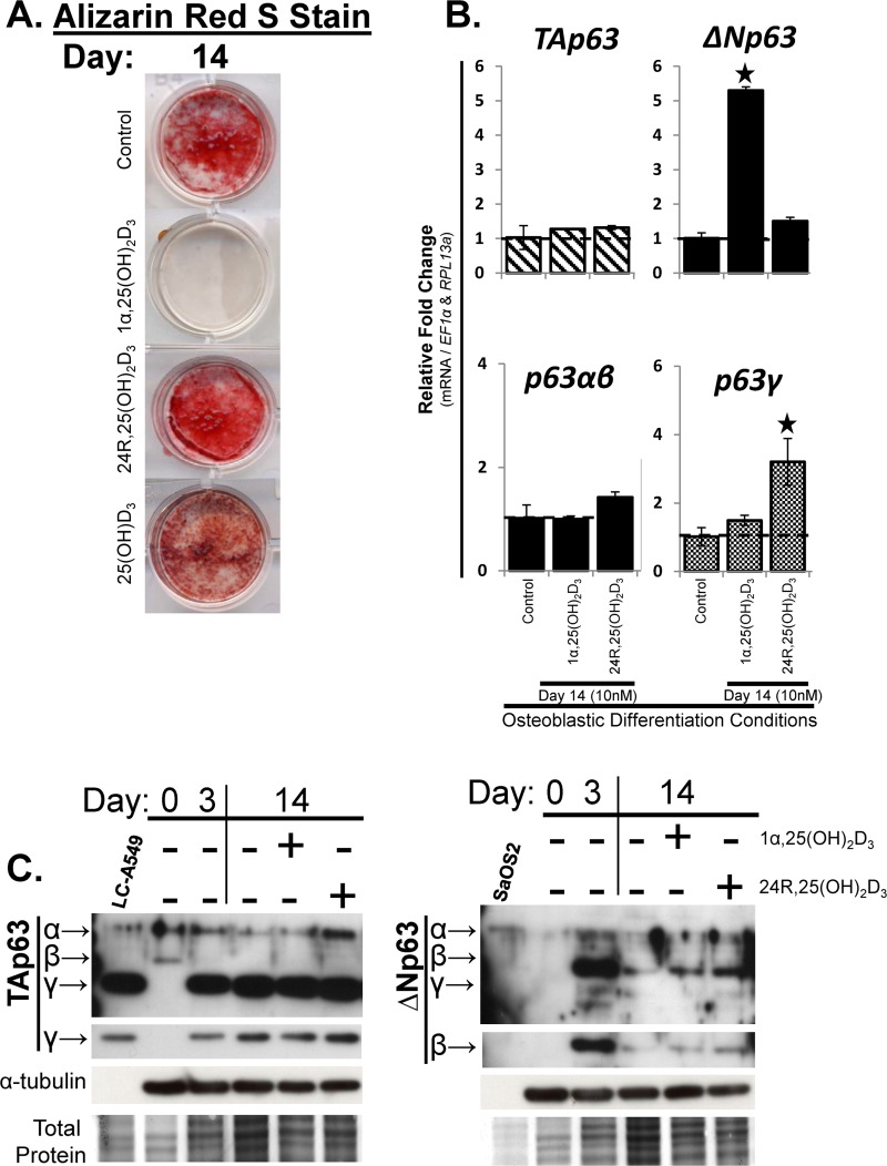
Fig 4. TAp63γ and ΔNp63β are the predominant p63 variants during osteoblastic differentiation of hMSC.
hMSC were seeded at 10,000 cells/cm2 under expansion conditions overnight then switched to osteoblastic differentiation conditions, without dexamethasone (Day 0). hMSC were treated with vitamin D3 metabolites 1α,25(OH)2D3 or 24R,25(OH)2D3 (10nM), or the vitamin D3 pro-hormone 25-hydroxyvitamin D3 (25(OH)D3) (10nM), starting at Day 0, and then re-treated every 3 days with media changes through Day 14. Control groups had no vitamin D3 treatments. Cells were fixed or harvested for RNA and protein at Days 3 and 14. A) Alizarin Red-S stain was used to determine Ca2+ mineralization. B) RT-qPCR analysis of p63 isoform (top panels, TA- and ΔNp63) and splice variant mRNA (bottom panels, p63α / β and p63γ) expression. C) Western blot analysis of p63 isoforms using antibodies detecting TAp63 variants (left panel, TAp63α, β, γ) or ΔNp63 variants (right panel, ΔNp63α, β, γ) during expansion conditions (Day 0) and under osteoblastic differentiation conditions (Days 3 and 14). Coomassie Blue = Total protein. Positive controls: TAp63: LC-A549 cells (positive for TAp63γ and low-expression of TAp63α protein) and ΔNp63; SaOS2 cells (positive for ΔNp63 variants). N = 3 independent experiments in triplicate. (*) p ≤0.05 compared to control (expanded, untreated hMSC). hMSC used were from a 7 and 22 year old male.

