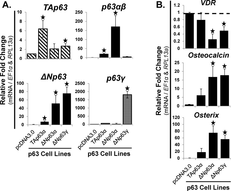
Fig 6. Stable overexpression of ΔNp63α and ΔNp63γ increased the mRNA expression of osteocalcin and osterix, and decreased VDR.
hMSC were transfected with pcDNA3.0-p63 vectors (7 days), followed by selection with G418 (3 weeks). The cells were then expanded and cloned over 2 months. Overexpression of TAp63β, TAp63γ and ΔNp63β caused a cell proliferation arrest; hence they were not available for long-term expansion and analysis. A) RT-qPCR analysis of p63 isoform (left panels, TA- and ΔNp63) and splice variant mRNA (right panels, p63α / β and p63γ) expression. RT-qPCR analysis demonstrated the relative level of overexpression of each of the p63 variants compared to empty vector (pcDNA3.0). B) RT-qPCR analysis of mRNA expression of osteoblastic differentiation markers: VDR (vitamin D3 receptor (binds 1α,25-dihydroxyvitamin D3)), Osteocalcin, and Osterix. Also of note, the overexpression of TAp63α, ΔNp63α, and ΔNp63γ did not alter the mRNA expression of runx2, osteopontin or the late stage osteoblastic gene, BSP. N = 3 independent experiments in triplicate. (*) p ≤ 0.05 compared to control (hMSC with empty vector, pcDNA3.0). hMSC used were from a 22 year old male.

