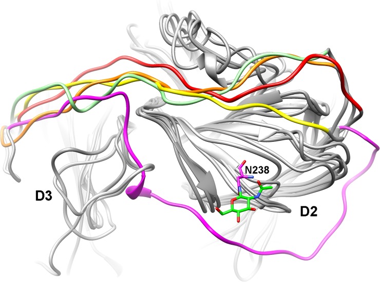
Fig 10. Comparisons of D2-D3 loop orientations.
Four bacterial laccases (PDB #2FQG, #1GSK, #2YAE and #4F7K) are superimposed with nLcc4. The D2-D3 loop in nLcc4 is threaded in direct proximity to the binding pocket; the D2-D3 loops in the other four bacterial laccases show the same orientation to connect D2 and D3 domains and pass near the N-terminus, where they cannot interact with any of the SBPLs. The D2-D3 loops from 2FQG, 1GSK, 2YAE, 4F7K and nLcc4 are colored orange, green, yellow, red and magenta, respectively.

