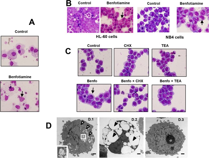
Fig 2. Benfotiamine induced cytoplasmic vacuolization in myeloid leukemic cells.
Primary leukemia cells BM-S-blasts (A) were cultured for 72 hours in the presence or absence of benfotiamine (100 μM) and subjected to Giemsa staining. Representative results of three independent experiments are shown. (B) HL-60 and NB4 cells were cultured for 72 hours in the presence of benfotiamine (100 μM) and then subjected to Giemsa staining. Arrows indicate the presence of cytoplasmic vacuoles. (C) HL-60 cells were cultured in the presence of 0.25 μg/ml of cycloheximide (CHX) or tetrathylammonium (TEA) for 2 hours, cultured for an additional 12 hours with or without 100 μM benfotiamine (Benfo) and stained with Giemsa. A representative result of three independent experiments is shown. (D) Transmission electron microscopic images of HL-60 cells treated with (D.1, D.2) or without (D.3) benfotiamine. The characteristic signs of paraptosis in benfotiamine-treated cells with massive vacuolization (open arrowheads) are visible. The inset in D.1 is a higher magnification showing mitochondria enlargement. Fig. D.2 shows megavacuoles occupying the cytoplasm of benfotiamine-treated HL-60 cells (closed arrowheads). Fig. D.3 shows untreated HL-60 cells with normal mitochondria architecture and rough endoplasmic reticulum. Bar = 1μm.

