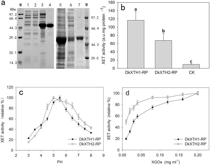
Fig 9. Expression and activity of recombinant XTH proteins.
(a) Proteins were separated on SDS–polyacrylamide gels and stained with Coomassie Blue. Lane 1, pET32a(+) control protein; lane 2, unbound protein (DkXTH1); lane 3, total protein (DkXTH1); lane 4, purified protein (DkXTH1); lane 5, total protein(DkXTH2); lane 6, unbound protein (DkXTH2); lane 7, purified protein (DkXTH2); and M, protein marks (Takara, Dalian, China). (b) In vitro XET assay of recombinant XTH proteins. The XET assay was performed by colorimetric method as described in Section 2.7. The empty vector pET32a(+) was used as the control. Columns with different letters are significantly different (LSD, P = 0.05) (c) The pH–rate profile of recombinant XTH proteins. (d) Dependence of XET activity of proteins on the concentration of XGOs. Vertical bars indicate standard errors of three replicates.

