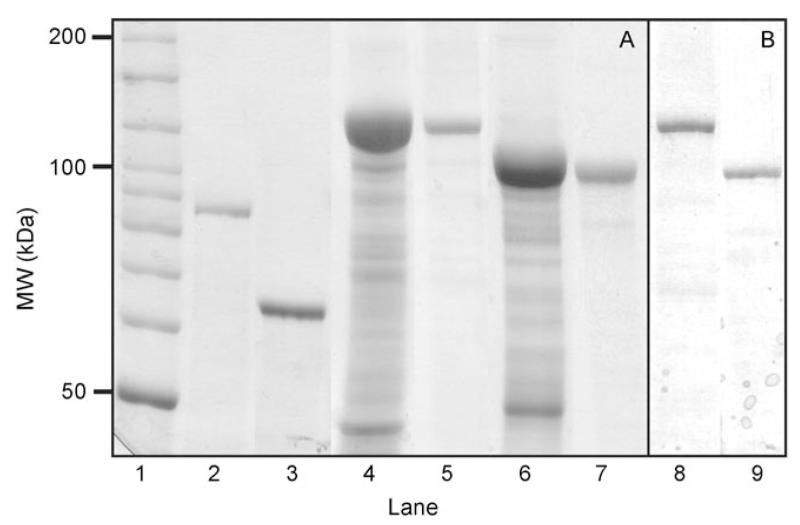
Figure 2.
A) SDS-PAGE analysis (8% gel) after Coomassie staining and B) Western-blot analysis using an antiRmlA antibody of biocatalytic proteins. Lane 1: Bench-mark ladder (Invitrogen); lane 2: purified C_SgsE (control); lane 3: purified G_SgsE (control); lane 4: raw extract of a C_SgsE.RmlA expression culture; lane 5: purified C_SgsE.RmlA; lane 6: raw extract of a G_SgsE.RmlA expression culture; lane 7: purified G_SgsE.RmlA; lane 8: purified C_SgsE.RmlA after Western blotting; lane 9: purified G_SgsE.RmlA after Western blotting. 20 μg (raw extracts) and 10 μg (purified proteins) of total protein were loaded on the gel for Coomassie staining; ≈2 μg were loaded for Western blotting.

