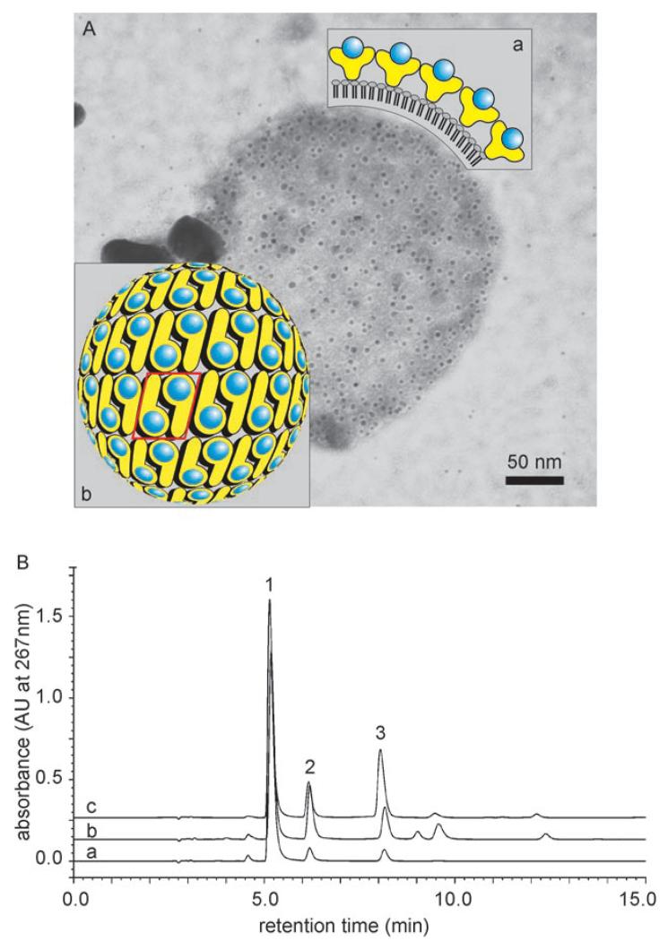
Figure 4.
A) Electron micrograph of an immuno gold-labeled (anti-RmlA antibody) and negatively stained liposome covered with a biocatalytic G_SgsE.RmlA monolayer. Inset: a) schematic cross section of a biocatalytic liposome, showing the surface display of the RmlA epitopes within the nanolattice; b) schematic representation of the p2 symmetry of the G_SgsE.RmlA nanolattice. Blue circles: biocatalytic RmlA epitopes (arbitrary positions); yellow symbols: G_SgsE carrier protein. One morphological unit of the nanolattice, corresponding to a G_SgsE.RmlA dimer, is boxed. B) Enzymatic activity of liposome-type biocatalyst was demonstrated by the formation of dTDP-d-glucose as analyzed by HPLC: a) sole RmlA; b) C_SgsE.RmlA, c) G_SgsE.RmlA 1: dTTP; 2: dTDP; 3: dTDP-d-glucose.

