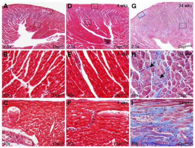
Figure 3.
Dsprul heart sections stained with Masson’s Trichrome. (A) 4 week old wild-type littermate control heart section. High magnification view of (B) left ventricular free wall (lower box in panel A) and (C) epicardium (upper box in panel A). (D) 4 week old homozygous mouse heart section. High magnification view of the (E) left ventricular free wall and (F) epicardium. (G) 34 week old homozygous, left ventricular free wall illustrating extensive fibrosis and a prominent fibrotic lesion. (H) High magnification view of left ventricular free wall fibrosis in aged homozygous mice (middle box in panel G). (I) High magnification view of a fibrotic lesion in the apex of the left ventricle (top, left box in panel G).

