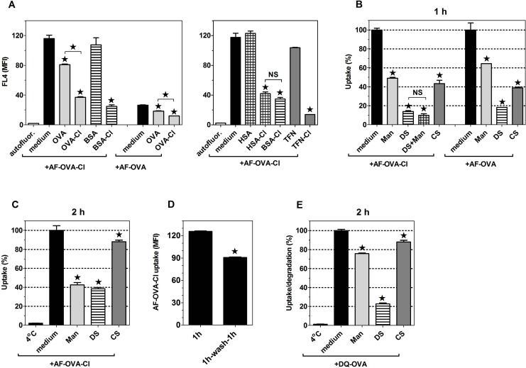
Fig 5. OVA and OVA-Cl bind to the same receptors, which are inhibited by mannan (Man), DS and CS and also shared with other HOCl-modified proteins and glycoproteins.
(A, B, C) BM-DC were pre-incubated for 20 min with 1 mg/ml of indicated unlabelled proteins (A), 6 mg/ml Man, 0.2 mg/ml DS or CS (B, C) before the same volume of double-concentrated solution of AF-OVA or AF-OVA-Cl was added to give the final concentration of 5 μg/ml and the incubation was continued for 1 h (A, B) or 2 h (C) in a cell culture incubator. Following washing, cell-associated fluorescence was quantified by flow cytometry. (D) BM-DC were incubated for 1 h with 5 μg/ml AF-OVA-Cl, washed and either directly assessed for antigen uptake or incubated for another 1 h in medium alone before the cell-associated fluorescence was measured. (E) Following pre-incubation with DS or Man, BM-DC were incubated for 1 h on ice with 20 μg/ml DQ-OVA. Unbound DQ-OVA was washed out and the cells were either directly assessed for DQ-OVA binding (“4°C”) or transfer to 37°C for 2 h before the measurement. Results shown are averages ± SEM of triplicates obtained in single experiments, each repeated at least 3 times with similar results. Statistical analysis was performed with ANOVA, followed by the Tukey-Kramer post-test (A-C, E) or with the Student’s t-test (D). *, p < 0.05; NS, non-significant.

