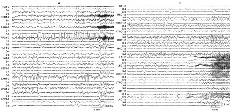
Fig 1. Examples of two depth electrode-recorded ictal EEG onset patterns from two different patients diagnosed with unilateral mesial temporal lobe epilepsy.
(A) Depth EEG recording displayed in a bipolar montage of a focal hypersynchronous ictal onset pattern consisting of low frequency (<2 Hz), high amplitude spikes in RPG1-2 that precedes the spread to RAH1-2 and REC1-2. This seizure spread to LPG1-2 and LAH1-2 1 min 21 sec after ictal onset (not shown). Total seizure duration was 2 min 56 sec. (B) Depth EEG recording of a low voltage fast ictal onset pattern, which compared to preceding EEG baseline, begins with low amplitude, high frequency (~33 Hz) activity nearly simultaneously in LEC1-2 and LA1-2. Note ictal activity appears in LOF3-4 and 5–6 within 5 sec and in RA1-2 11 sec after ictal onset. Total seizure duration was 1 min 10 sec. Depth electrodes labels and anatomical location as follows: R (right)/L (left) A (amygdala), AC (anterior cingulate), AH (anterior hippocampus), EC (entorhinal cortex), OF (orbitofrontal cortex), PG (posterior parahippocampal gyrus), STG (superior temporal gyrus), and TPO (temporal-parietal-occipital junction); numbers refer to electrode contacts 1 (distal, medial surface) to 6 (proximal, lateral surface).

