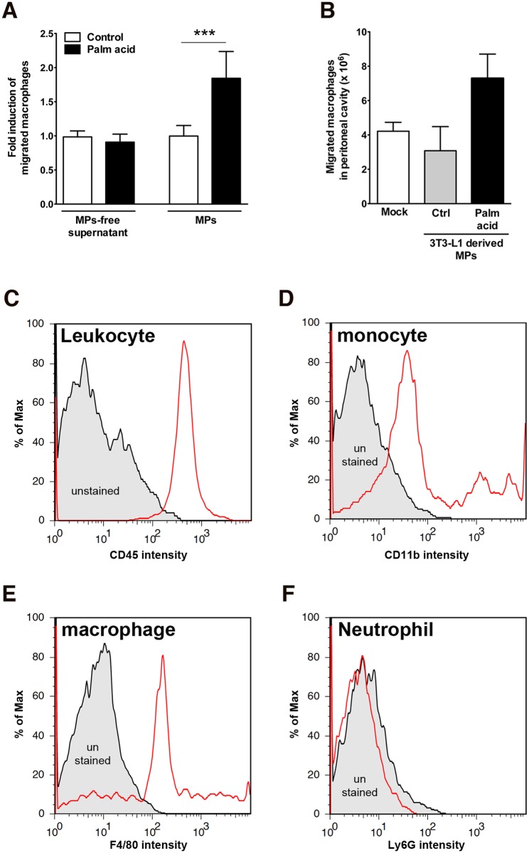
Fig 4. Adipocyte-derived MPs mediates attraction of macrophages in vitro and in vivo.
(A) In vitro chemotaxis assay of MPs-free supernatant and MPs from adipocytes treated with palmitic acid. Values represent mean ± S.D. ***P < 0.001 compared to controls. (B-F) C57BL/6 mice were injected intraperitoneally with 1x10⁶ adipocyte-derived MPs (isolated from 3T3-L1 adipocytes treated with 0.5 mM palmitic acid or without palmitic acid) or controls (n = 3 each group). (B) Four days post injection infiltrated cells were isolated from peritoneal cavity by lavage and counted. (C-F) flow cytometry analysis of infiltrated cells. The infiltrated cells were stained by CD45 (leukocyte common antigen) (C), CD11b (monocytes) (D), F4/80 (macrophages) (E), or Ly6G (neutrophils) (F). Values represent mean ± S.E.M. (D) In vivo macrophages migration. C57BL/6 mice were injected intraperitoneally with 1x10⁶ palmitic acid-derived MPs or vehicle alone (n = 5 per group). Three days post injection; macrophages were isolated from peritoneal cavity by lavage. Number of macrophages present in peritoneal cavity was counted. Values represent mean ± S.E.M.

