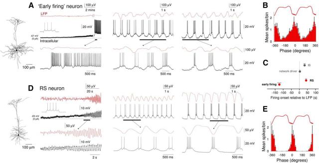
Figure 9.
Comparison of the development of UP states in “early firing” and non-“early firing” RS neurons following cholinergic receptor activation. A, Simultaneous recording of LFP and intracellular activity of an “early firing” neuron. Left (onset), Following bath application of CCH, the neuron is strongly depolarized and starts to exhibit continuous action potential firing. At this point, there is no oscillatory activity in the LFP. The underlined section is expanded below as indicated. Inset top, The response of this neuron to a depolarizing current step. Middle (early), After a few minutes, oscillatory activity is established in the LFP. This is gradually accompanied by the appearance of hyperpolarizing excursions in the membrane potential of the “early firing” cell. Right (late), Following prolonged application of CCH, activity in the “early firing” neuron on each oscillation cycle takes on a characteristic appearance consisting of a ramp-like depolarization and buildup of action potential firing, followed by a more abrupt hyperpolarization. The reconstructed morphology of this neuron is shown to the far left. B, Average spike timing histogram (mean ± SEM) of 6 “early firing” neurons. C, Quantification (mean ± SEM) of the delay between the onset of the oscillation in the LFP and onset of firing for “early firing” (n = 11) and non-“early firing” RS (n = 10) neurons; “network driver” and conventional IB neurons are also shown for comparison. D, Simultaneous recording of LFP and intracellular activity of a non-“early firing” RS neuron. Left (onset), Following bath application of CCH, the neuron is moderately depolarized and starts to exhibit rhythmic EPSP complexes in synchrony with the emergent oscillation in the LFP. The underlined sections are expanded below as indicated. Inset top, The response of the neuron to a depolarizing current step. Middle (early), After a few minutes, EPSP complexes start to be crowned by one or two action potentials. Right (late), Following prolonged application of CCH, the neuron exhibits full-blown UP states with a typical step-like appearance. The reconstructed morphology of this neuron is shown to the far left. E, Average spike timing histogram (mean ± SEM) of 6 non-“early firing” RS neurons.

