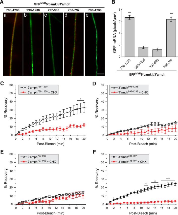
Figure 4.

A 60 nt segment of amphoterin 3′-UTR is sufficient for axonal localization in DRG neurons. A, Representative exposure-matched FISH/IF images for axons of neurons transfected with indicated GFPMYR-5′camkII/3′-amph constructs. GFP mRNA is shown in red and NF protein in green (antisense probes are shown in Aa–Ad and sense cRNA probe is shown in Ae). Quantifications of GFP RNA FISH signals in DRG axons across multiple experiments are shown in B as average ± SEM (n ≥ 30 over three independent experiments; ***p ≤ 0.001 by Student's t test). Scale bar, 10 μm. C–F, Postbleach signals from FRAP studies for distal axons of DRG neurons transfected with the same GFPMYR constructs as in A are shown. In each case, the ROI was at least 400 μm from the cell body. Average signal intensity normalized to prebleach and postbleach signal for each experiment is shown; error bars indicate the SEM of normalized data (n ≥ 7 over at least 3 independent experiments). CHX, Cultures pretreated with 150 μg/ml cycloheximide for 20 min before photobleaching (n ≥ 4 over at least 3 independent experiments; *p ≤ 0.05, **p ≤ 0.01, and ***p ≤ 0.001 for indicated time points compared with t = 0 min postbleach by repeated-measures ANOVA with Bonferroni post hoc comparisons).
