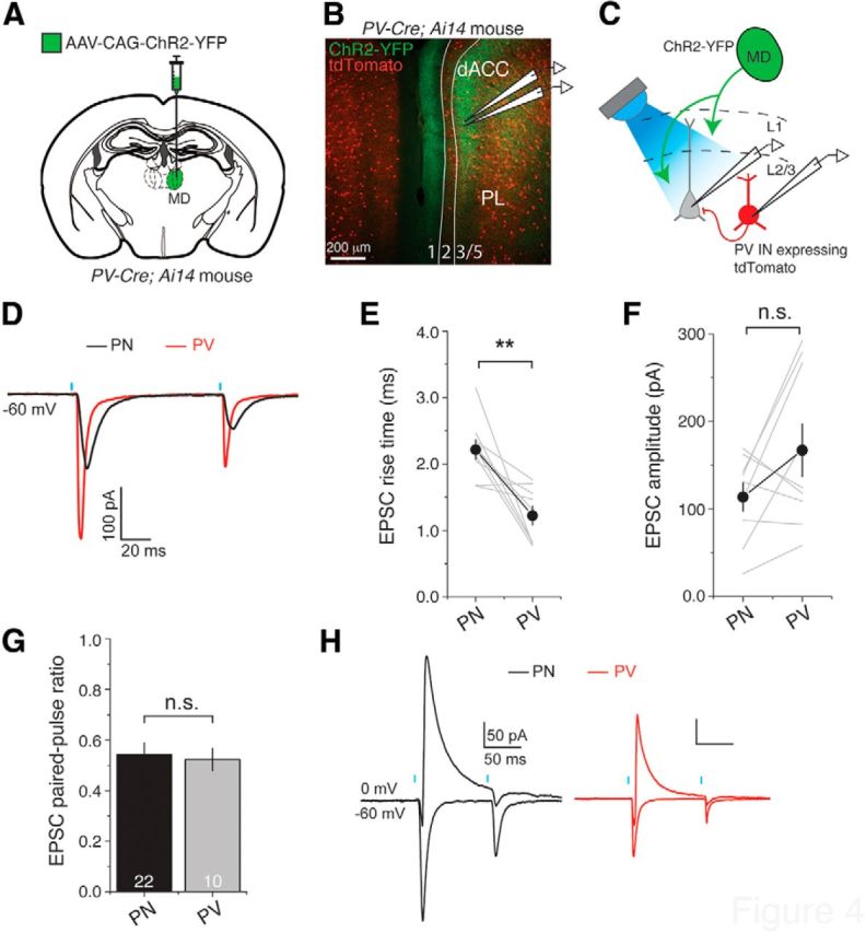
Figure 4.

MD neurons directly excite PV interneurons in layer 3 of dACC. A, Schematic of the experimental approach. B, Representative image of a brain section from a PV-Cre;Ai14 mouse, in which the MD was injected with AAV-CAG-ChR2-YFP. The ChR2-YFP-expressing axons originating from the MD terminate in layers 1, 3, and 5 in dACC. PL, Prelimbic cortex. C, Schematic of the recording configuration. D, EPSCs evoked by photostimulation of inputs from the MD were sequentially recorded in pairs of neighboring PV INs and PNs in dACC. E, The rise time of EPSCs was faster in PV INs than in PNs. **p < 0.005 (paired t test). F, G, The amplitude (F) or paired-pulse ratio (100 ms interpulse interval) (G) of EPSCs in PV INs was not significantly different from that in neighboring PNs. n.s., Not significant. Numbers in bar graph indicate the number of cells recorded. H, PV INs also receive feedforward inhibition. The MD-driven feedforward IPSCs onto PV INs or PNs showed strong paired-pulse depression (100 ms interstimulus interval). Data are mean ± SEM.
