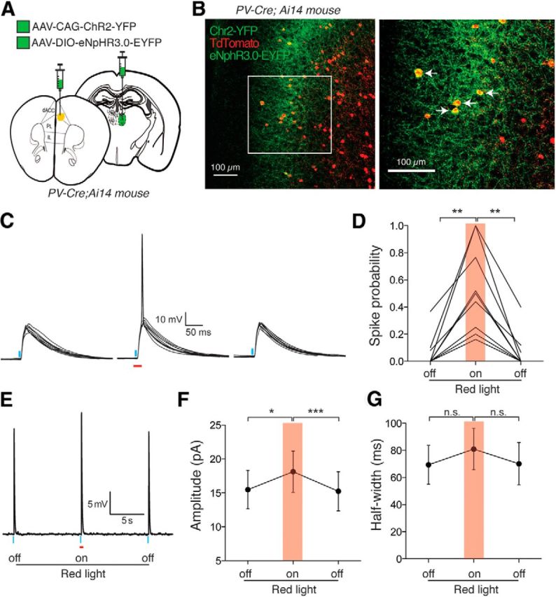
Figure 9.

PV IN-mediated feedforward inhibition controls PN spike output in the dACC. A, Schematic of injection. AAV-CAG-ChR2-YFP was injected into MD while AAV-DIO-eNpHR3.0-GFP was injected into dACC of PV-Cre;Ai14 mice in which PV INs are labeled by Tdtomato. B, Labeling of ChR2-YFP+ axons originating from MD, Tdtomato+ PV INs, and eNpHR3.0-EYFP-infected PV INs in L3 of the dACC. Arrows indicate neurons in which eNpHR3.0-EYFP and Tdtomato were coexpressed. C, Sample PSP traces recorded from an L3 PN in response to blue light activation (0.5 ms; blue bars) of MD axons (ChR2+) in the presence or absence of red light illumination (20 ms; red bar) to inhibit local PV INs (eNpHR3.0+). Red light was triggered 5 ms before the onset of the blue light pulse. D, Silencing PV INs reversibly increased ChR2-evoked spike probability in L3 PNs. **p < 0.01 (one-way repeated-measures ANOVA followed by Tukey's test). E, Averaged subthreshold PSP sample trace, showing the effect of red light illumination. F, Silencing PV INs reversibly increased the peak amplitude of ChR2-evoked subthreshold PSPs in L3 PNs. *p < 0.05, ***p < 0.001 (one-way repeated-measures ANOVA followed by Tukey's test). G, Silencing PV INs did not significantly alter the half-width of ChR2-evoked subthreshold PSPs in L3 PNs. n.s., Not significant (one-way repeated-measures ANOVA). Data are mean ± SEM.
