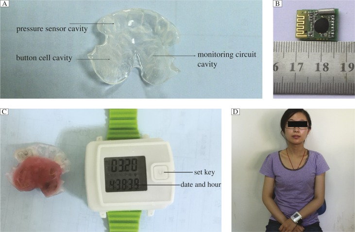
Fig. 2. Mini wireless biofeedback device.
A: The margin of the biofeedback splint extends to the lingual surface of the bilateral maxillary second premolars. The cavity design for placement of the pressure sensor, monitoring circuit and button cell. B: Mini-monitoring circuit (18 mm×16 mm×5 mm). C: A maxillary biofeedback splint with pressure sensor, monitoring circuit and button cell embedded(left), a watch style vibration device (right). D: A bruxer with the maxillary biofeedback splint for monitoring and the wireless vibration device. Use of photo was permitted by the study subject.

