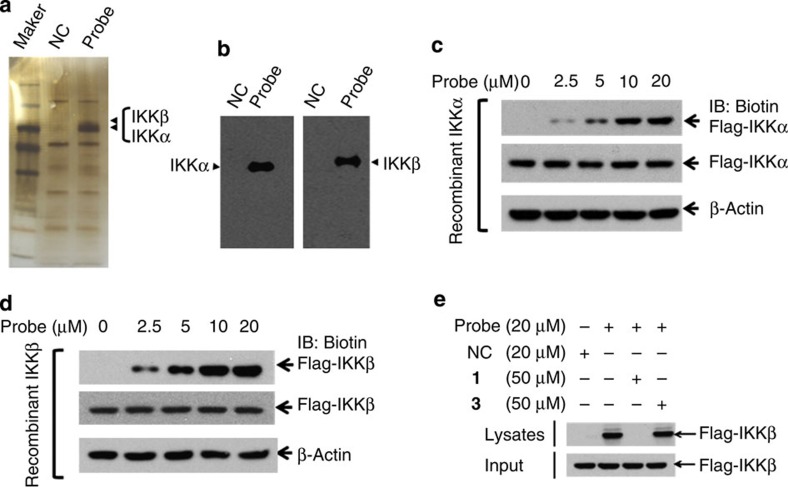
Figure 4. Ainsliadimer A (1) directly targets IKKα and IKKβ.
(a,b) The 293T cell lysates were incubated with Probe or NC at 4 °C overnight, the lysates were used for streptavidin-agarose pull-down assays, and the precipitates were resolved by SDS–PAGE, followed by silver staining. The indicated bands were excised and analysed via mass spectrometry (a), or detected by western blotting for IKK proteins as indicated (b). (c,d) The recombinant IKKα and IKKβ proteins were incubated with Probe or NC for 1 h at 37 °C, followed by immunoblotting with biotin (upper band) or flag (lower band). (e) The recombinant IKKβ protein was incubated with Probe in the absence or presence of a twofold excess of unlabelled ainsliadimer A (1) or 3 for 1.5 h at 37 °C, and the mixtures were blotted for biotin or Flag. All experiments were repeated at least three times, and similar results were obtained each time.

