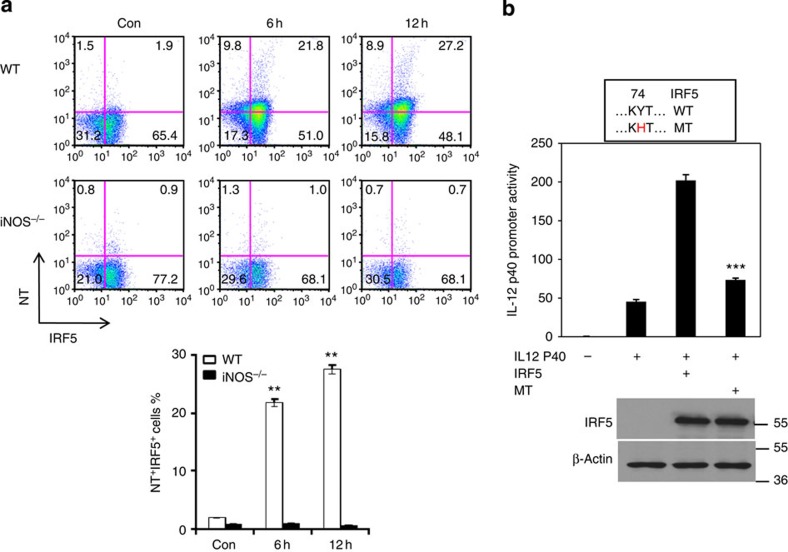
Figure 6. NO induces nitration of tyrosine residues of IRF5 protein in macrophages.
(a) WT or iNOS−/− mice were injected (i.p.) with LPS (200 μg/mouse) for 6 or 12 h. Mice were then killed and spleen cells were prepared. The cells were stained for IRF5 and nitrotyrosine and analysed by flow cytometry. Representative FACS dot plots gated on CD11b+ cells, and the percentages of IRF5 and nitrotyrosine-positive CD11b+ cells are shown. Each bar represents mean±s.d. from three independent experiments, unpaired Student’s t-test, **P<0.01, versus iNOS−/− cells. (b) The 293T cells were transfected with an IL-12 p40 promoter reporter construct and wild-type IRF5 or mutant IRF5Y74H plasmids for 30 h. Luciferase assays were performed, and luciferase activities were normalized to β-galactosidase activity. In addition, IRF5 protein expression was analysed by western blotting. Each bar represents mean±s.d. from three independent experiments, unpaired Student’s t-test, ***P<0.001, versus cells transfected with WT IRF5 plasmid.

