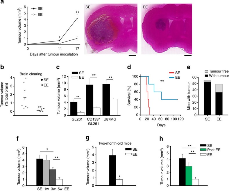
Figure 1. Exposure to EE reduces glioma growth and prolongs survival.
(a) The mean tumour volumes at 11 and 17 days after implantation of glioma cells into the striatum of C57BL/6 mice, housed in SE or EE, as indicated, from at least 11 mice per conditions. *P<0.05, **P<0.01, Student’s t-test. Representative coronal brain sections are shown on the right for each condition at 17 days. Scale bar, 1 mm. (b) Quantification of tumour volumes under the same experimental conditions as in a, at 17 days, with the brain-clearing technique (n=7–8; **P<0.001 Student’s t-test). Representative Movies are shown as Supplementary Material.(c) The mean tumour volumes (±s.e.m.) at 17 days after implantation of GL261, purified CD133+ GL261 or U87MG glioma cells into the striatum of C57BL/6, or SCID mice housed in SE or EE, as indicated; seven animals per condition. **P<0.01, Student’s t-test. (d) Kaplan–Meier survival curves of SE (red) and EE (blue) GL261 glioma-bearing mice (n=5 per group; log-rank test **P<0.01). (e) Tumour resistance in SE and EE mice expressed as total number of mice with (black) and without (grey) brain tumour 17 days after GL261 transplantation. (f) ‘Dose dependency’ of EE exposure on tumour volume (n=7 per group; *P<0.05; **P<0.01, one-way ANOVA. (g) Effect of SE or EE housing on 2-month-old mice transplanted with glioma (n=5 per group; *P<0.05, Student’s t-test). (h) Effect of housing in SE and EE before (EE, for 5 weeks) or after tumour transplantation (post-EE, for 17 days) on tumour volume (all mice analysed 17 days after tumour transplantation). Data are from at least five mice per group. **P<0.01, Student’s t-test.

