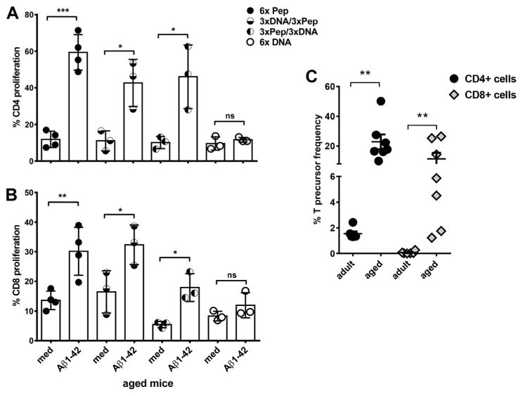
Fig. 4.

Analyses of T cell proliferation in adult and aged mice with a CFSE proliferation assay. Mouse splenocytes were labeled with CFSE and cultured for 5 days with medium or Aβ42 peptide. After staining with antibodies for CD4 and CD8, CFSE dilution was measured following gating on the respective T cell populations by flow cytometry. Proliferation was measured from triplicate wells for each of the mice analyzed. One circle represents the proliferation found for 1 individual mouse. In A, CD4 proliferation is compared for the differently immunized aged mouse groups (mean values and SEM). In B, CD8 proliferation is shown (mean values and SEM). Using the FCS3 express software, CD4 and CD8 precursor frequencies were calculated based on numbers and daughter generations of divided cells for adult and aged Aβ42 peptide immunized mice (C). Results shown are representative for 3 similar performed experiments. *** (p<0.0005), ** (p<0.005),* (p<0.05), ns (p>0.05). Abbreviations: CFSE, carboxyfluorescein succinimidyl ester; SEM, standard error of the mean.
