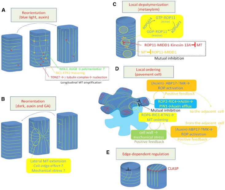
FIGURE 1.
Regulation of cortical microtubule rearrangements. (A) Reorientation from transverse to longitudinal. (B) Reorientation from longitudinal to transverse. (C) Local depolymerization in xylem vessel cells. (D) Local ordering in leaf pavement cell. (E) Cell edge-dependent regulation by CLASP protein. Green lines indicate cortical microtubules (A–E). Red lines in (D) indicate actin microfilaments. MT, microtubule; GA, gibberellic acid.

