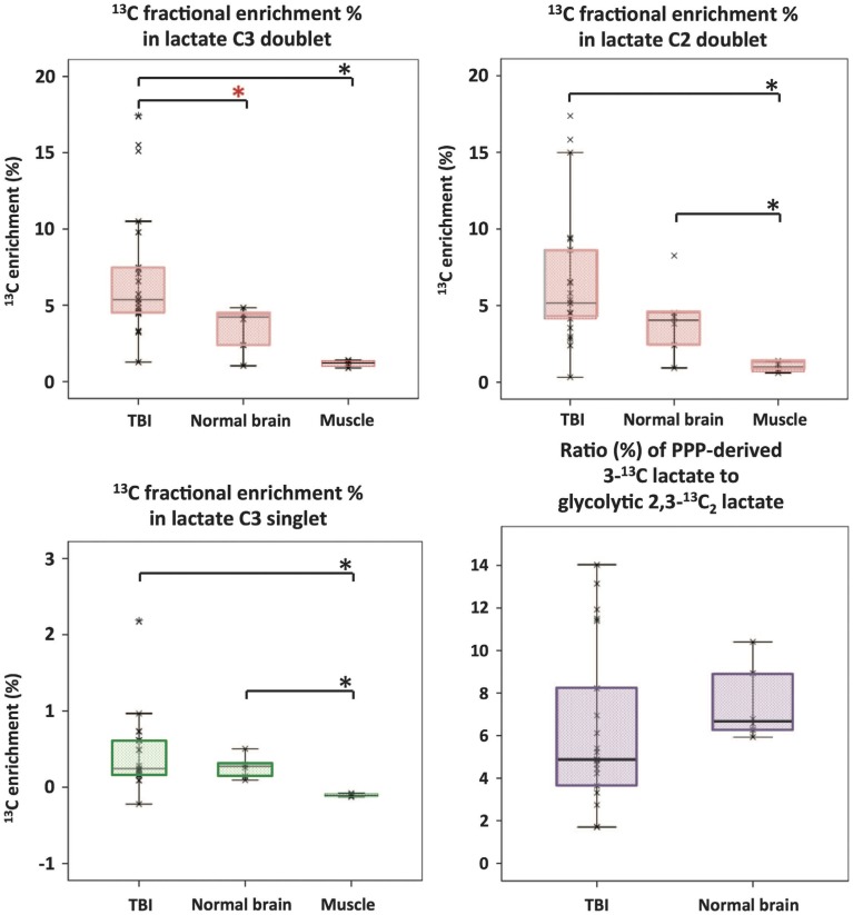
Figure 7.
Microdialysate NMR measurements of 13C labeling: results from perfusion for 24-h- (brain: TBI or “normal”) or 8-h perfusion (muscle) with 1,2-13C2 glucose (4 mmol/L). Red asterisks denote P < 0.01 for TBI vs. “normal” brain (Mann–Whitney); other comparisons asterisked in black denote P < 0.05. Individual data points are shown by × symbols. Number of patients: 15 TBI, six “normal” brain, and four muscle. Originally published by Jalloh et al. (2015) in J Cereb Blood Flow Metab 35: 111–120, and reproduced with permission of Nature Publishing Group.

