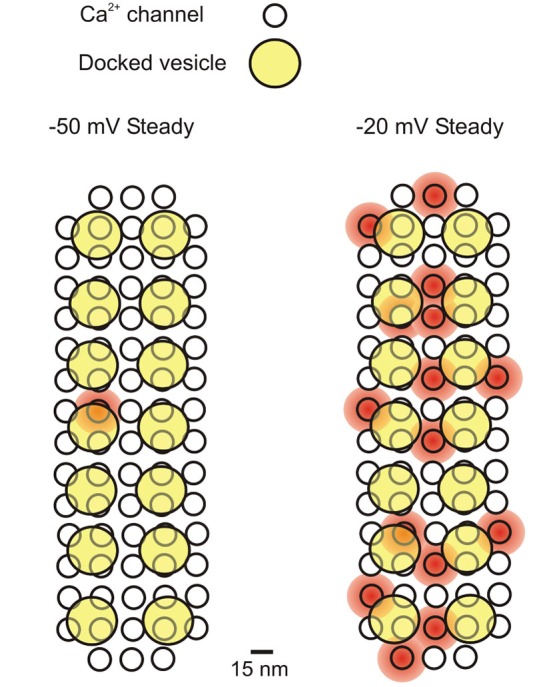
Figure 1.

Schematic representation of the Ca2+ influx-exocytosis coupling for a presynaptic active zone in a mature IHC. Ca2+ channels simultaneously open at −50 mV and −20 mV (during a 500 ms depolarizing step from −70 mV), representing the average Po found in cell-attached recordings: 0.01 and 0.21 at −50 and −20 mV, respectively (Zampini et al., 2013). It is assumed that each presynaptic site (active zone) contains 74 Ca2+ channels (see text) and 14 docked vesicles (Wong et al., 2014). The diameter of Ca2+ channels is ~15 nm (Wolf et al., 2003), while that of vesicles is ~40 nm (Khimich et al., 2005). Placing 74 Ca2+ channels closely packed (7.5 nm minimum distance) gives a presynaptic area of ~0.033 μm2, which is consistent with the most recent EM estimates (0.066 μm2; Khimich et al., 2005) and STED microscopy (0.11 μm2 and 0.097 μm2 for apical and middle cochlea IHCs, respectively: Meyer e al., 2009; 0.034 μm2 for mature IHCs: Wong et al., 2014). The shaded red circular areas indicate a 20 nanometer distance of cytosolic Ca2+ diffusion from the central pore of the Ca2+ channel when it is open. Kim et al. (2013) calculated that a Ca2+ channel current of 0.5 pA (we estimated 0.8 pA at −50 mV and 0.4 pA at −20 mV; Zampini et al., 2013) will produce a [Ca2+]i of 350 μM near the channel mouth, which would decrease to 50–100 μM in a presynaptic region including three or four docked vesicles. Here we assumed that Ca2+ entering from each channel can reach at least one docked vesicle. If all Ca2+ channels showed an analogous gating behavior, then on average 1 (out of 74) Ca2+ channels (each with a Po of 0.01) would be always open at −50 mV at the presynaptic site, and 15 (each with a Po of 0.21) at –20 mV.
