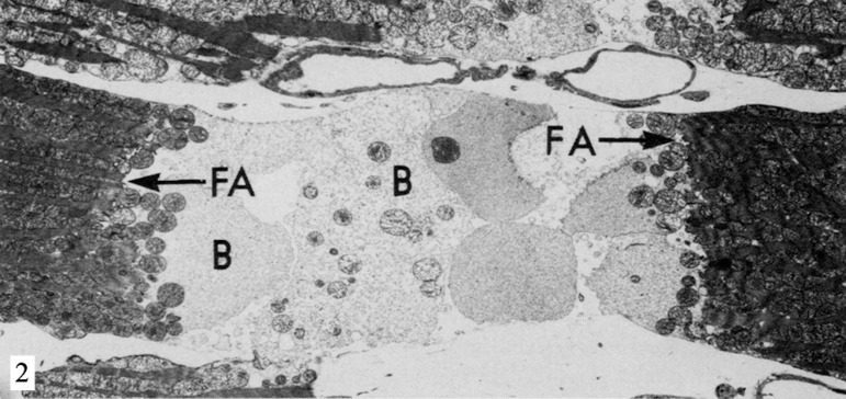
Fig. 2.
Electron micrograph of rat heart after 5 minutes in calciumfree perfusion, followed by 15 minutes with the addition of DNP at the first perfusate. The sarcomeres are contracted herein, pulling the fascia adherens (FA) and causing damage to the cell membrane of the myocyte in the nexus area. The cytosol is exteriorized in the form of blebs (B) in the intercellular region. Reproduced from Ganote et al., 1985[23]

