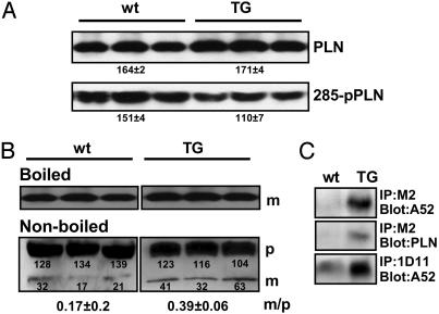
Fig. 4.
Properties of PLN in NF-SLN TG mice. (A) Western blot analyses of ventricular samples to detect PLN and phospho-PLN (285-pPLN) and PLN from wt and TG hearts. Numbers represent the mean ± SEM in arbitrary densitometric units. Note the reduced level of PLN phosphorylation in TG samples. (B) Monomer/pentamer ratio (m/p) of PLN in TG hearts. Boiled or nonboiled microsomal fractions were subjected to immunoblotting with anti-PLN antibody. p, Pentameric PLN; m, monomeric PLN. (C) Physical interaction of PLN with SERCA2a in TG hearts. Microsomal fractions were subjected to immunoprecipitation with anti-PLN antibody, 1D11, or anti-FLAG antibody, M2. Precipitates were separated by SDS/PAGE, and SERCA2a or PLN were detected by immunoblotting.

