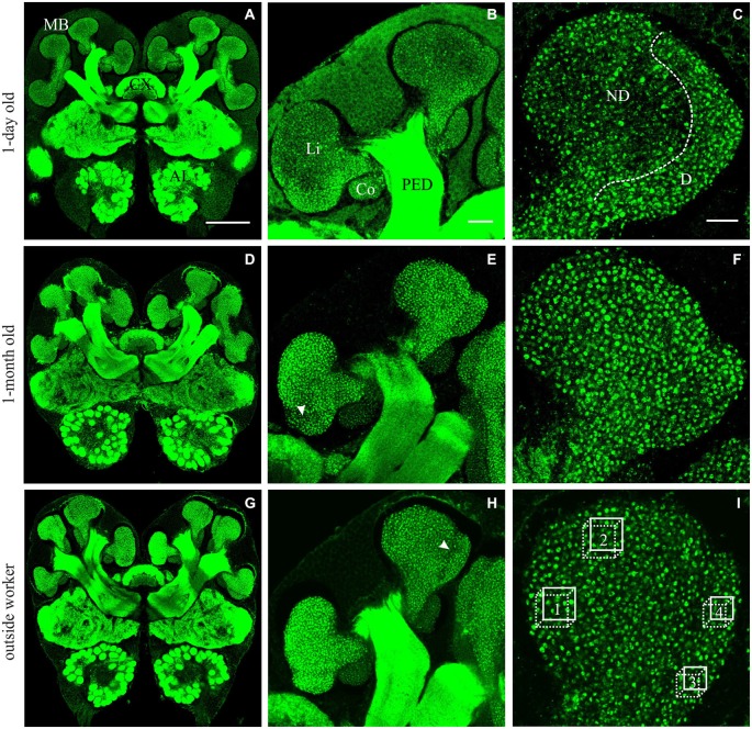
Figure 1.
Immunofluorescence staining with anti-synapsin in brains from A. ambiguus workers of different ages: examples of 1-day old (A–C), 1-month old (D–F), and from outside-workers of unknown age (G–I). (A,D,G) Frontal overview of brains in a central plane. mushroom bodies (MBs), central complex (CX) and antennal lobes (AL) are indicated. (B,E,H) Comparison of the staining of MB calyces at different ages. Overview of the left medial calyx: lip (Li), collar (Co) and peduncle (PED). (C,F,I) Higher magnification of the lip calycal region. In 1-day old ants, the lip appeared differentially stained. The non-dense (ND) lip presented lower number of synapsin-immunoreactive (IR) boutons compared with the dense (D) lip. In older ants, in which the boutons clearly appeared as well defined structures, this differentiation was not obvious and two subregions could be defined by an unstaining band between them—as was previously reported for other ant species (Gronenberg, 2001)–, indicated by the arrow in (E,H). In (I), boxes 1–2 and 3–4 indicate the volumes used for quantification of MGs in the ND and D subregions of the MB calyces, respectively. Scale bars: in (A) (valid for A,D,G): 100 μm; in (B) (valid for B,E,H): 20 μm; in (C) (valid for C,F,I): 10 μm.

