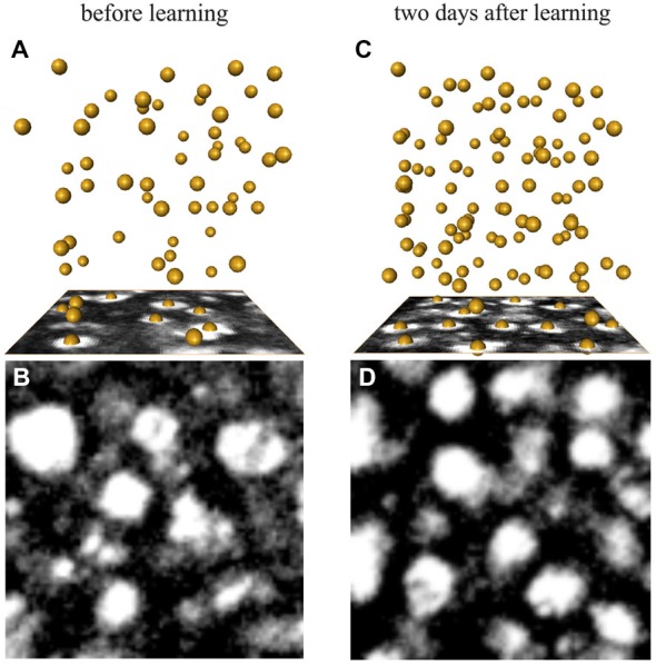
Figure 6.

Examples of synapsin-IR bouton quantification in the ND lip (A,B) before and (C,D) 2 days after learning. (A,C) Three-dimensional reconstruction of the position of the boutons visualized by AMIRA in a 1000 μm3-volume (86 × 86 voxel). Each yellow sphere marks the center of a bouton. (B,D) Single confocal image of a 10 × 10 μm2 (86 × 86 voxel) synapsin-stained area in the ND lip.
