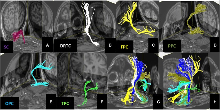
Figure 4.
3D views of the (A) Spinocerebellar, (B) Dentate-Rubro-Thalamo-Cortical, (C) Fronto-Ponto-Cerebellar, (D) Parieto-Ponto-Cerebellar, (E) Occipito-Ponto-Cerebellar, (F) Temporo-Ponto-Cerebellar pathways. All these cerebellar pathways are illustrated together with corticospinal tract (dark blue) in panels (G,H).

