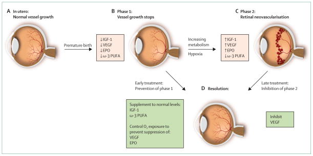
Figure 1. Progression of retinopathy of prematurity.
(A) Oxygen tension is low in utero and vascular growth is normal. (B) Phase 1: after birth until roughly 30 weeks postmenstrual age, retinal vascularisation is inhibited because of hyperoxia and loss of the nutrients and growth factors provided at the maternal–fetal interface. Blood-vessel growth stops and as the retina matures and metabolic demand increases, hypoxia results. (C) Phase 2: the hypoxic retina stimulates expression of the oxygen-regulated factors such as erythropoietin (EPO) and vascular endothelial growth factor (VEGF), which stimulate retinal neovascularisation. Insulin-like growth factor 1 (IGF-1) concentrations increase slowly from low concentrations after preterm birth to concentrations high enough to allow activation of VEGF pathways.
(D) Resolution of retinopathy might be achieved through prevention of phase 1 by increasing IGF-1 to in-utero concentrations and by limiting oxygen to prevent suppression of VEGF; alternatively, VEGF can be suppressed in phase 2 after neovascularisation with laser therapy or an antibody. EPO=erythropoietin. ω-3 PUFA=ω-3 polyunsaturated fatty acids. Adapted from reference 33, by permission of the Association for Research in Vision and Ophthalmology.

