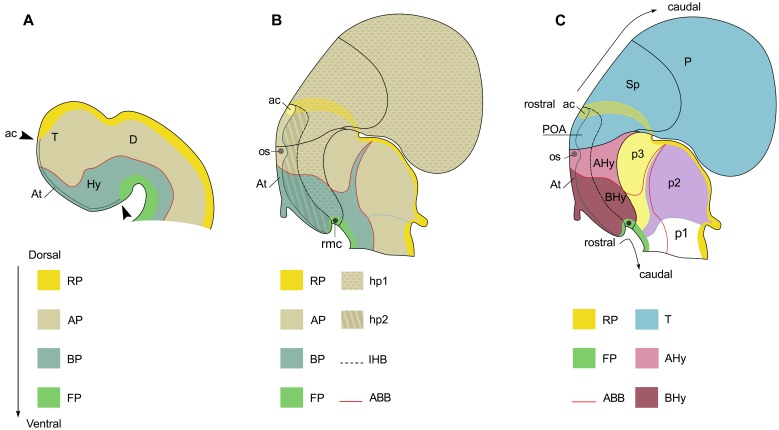
FIGURE 2.
Squematic representations of the prosencephalon of early (A) and late (B,C) mouse embryo to show correspondence of longitudinal and tranverse domains in the secondary prosencephalon under the updated prosomeric model. Domains in (A) are illustrated according to Figure 1.1C in Martínez et al. (2012). Domains in (B,C) are illustrated according to Figure 8.5B in Puelles et al. (2012). (A) Longitudinal domains in early embryos. The arrowheads mark both the dorso-caudal and ventro-caudal limits of the acroterminal territory (At). This territory is considered the rostral-most domain of the neural tube. The dorso-caudal limit of the At can be identified caudal to the anterior commissure. (B) Longitudinal and transverse organization in late embryos. (C) Segmental organization of the secondary prosencephalon according to the prosomeric model. For abbreviations, see list.

