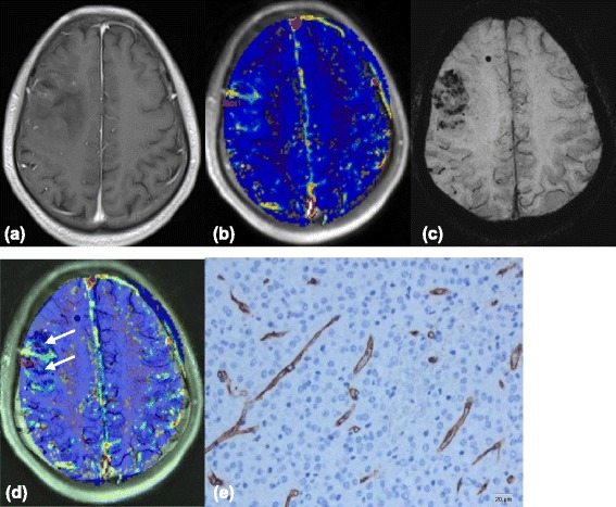
Figure 2.

Images(a-d) of a 26-year-old man with right frontal low-grade Oligodendroglioma. (a) Contrast-enhanced T1-weighted image shows fair enhancement of the tumor. (b) Ktrans map shows mild increased Ktrans values within the tumor, relative to the normal brain tissue. (c) Multiple dotlike ITSS are shown in the SWI. (d) Coregistered image of Ktrans and SWI shows that regions of the highest value of Ktrans does not correspond with areas of attenuated prominent ITSS(arrows) in the same segment. (e) Representative immunohistochemical staining(CD34, Original magnification,×200) shows rather abundant mirovessels with small VD.
